- English
- French
- German
- Portuguese
- Spanish
- Russian
- Japanese
- Korean
- Arabic
- Greek
- German
- Turkish
- Italian
- Danish
- Romanian
- Indonesian
- Czech
- Afrikaans
- Swedish
- Polish
- Basque
- Catalan
- Esperanto
- Hindi
- Lao
- Albanian
- Amharic
- Armenian
- Azerbaijani
- Belarusian
- Bengali
- Bosnian
- Bulgarian
- Cebuano
- Chichewa
- Corsican
- Croatian
- Dutch
- Estonian
- Filipino
- Finnish
- Frisian
- Galician
- Georgian
- Gujarati
- Haitian
- Hausa
- Hawaiian
- Hebrew
- Hmong
- Hungarian
- Icelandic
- Igbo
- Javanese
- Kannada
- Kazakh
- Khmer
- Kurdish
- Kyrgyz
- Latin
- Latvian
- Lithuanian
- Luxembou..
- Macedonian
- Malagasy
- Malay
- Malayalam
- Maltese
- Maori
- Marathi
- Mongolian
- Burmese
- Nepali
- Norwegian
- Pashto
- Persian
- Punjabi
- Serbian
- Sesotho
- Sinhala
- Slovak
- Slovenian
- Somali
- Samoan
- Scots Gaelic
- Shona
- Sindhi
- Sundanese
- Swahili
- Tajik
- Tamil
- Telugu
- Thai
- Ukrainian
- Urdu
- Uzbek
- Vietnamese
- Welsh
- Xhosa
- Yiddish
- Yoruba
- Zulu
Early Clinical Effect Of Tantalum On Femoral Head Necrosis
2024-01-05 18:00:06
1. Biological basis of tantalum application
Insoluble tantalum salt is not absorbed by human body after oral or local injection, and the amount of soluble tantalum salt absorbed by gastrointestinal tract is very small. Once tantalum enters the human body, phagocytes are the main carriers responsible for the removal of tantalum. Phagocytes in the body can survive after 1h of contact with tantalum dust without cell degeneration, and are only accompanied by a significant increase in glucose oxidation. Under the same conditions, silica dust can cause severe cytoplasmic degeneration and death of phagocytes, indicating that tantalum is cytotoxic. In 1940, pure tantalum was first used in orthopedic medicine, and most reports showed that tantalum metal as human implants had no adverse reactions.
2. Medical application of tantalum
2.1 Tantalum wire
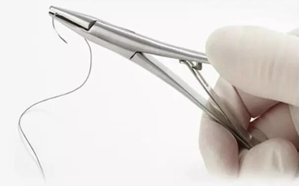
Tantalum is so malleable that it can be made as fine as or finer than human hair. Tantalum wire, as a surgical suture line, has the advantages of simple sterilization, small stimulation and high tension resistance, but it also has the disadvantages of not easily knotting. Tantalum wire can be used for suturing bones, tendons, fascia, as well as for reducing tension sutures or internal dental fixation. It can also be used as sutures for visceral surgery or inserted into artificial eyeballs. Tantalum wire can even replace tendons and nerve fibers. Xu hao et al. reported 33 cases of patella fractures of various types treated with tantalum wire band internal fixation, and followed up for 5 months to 6 years after the operation. Except 2 cases of mild traumatic arthritis, the other 31 cases achieved good efficacy without complications.

2.2 Tantalum sheet
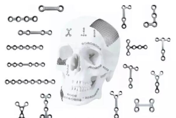
Tantalum metal can be made into tantalum chips of various shapes and sizes, which can be implanted according to the needs of various parts of the human body, such as repairing and sealing the cracks and defects of fractured cranium and limbs. After the head is fixed with tantalum, the skin is grafted from the legs. After a period of time, the grafted skin grows so well that it is hardly visible.

2.3 Tantalum scaffolds
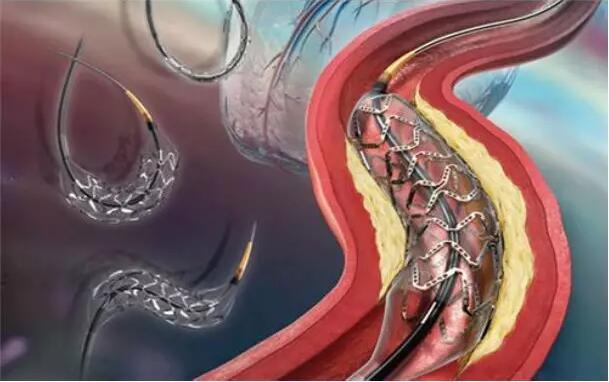
The tantalum wire can be woven into a mesh balloon dilating stent, which can be clearly seen under X-ray and is very convenient for monitoring and follow-up. Its long - term retention in the body without fracture and corrosion. Due to its good flexibility, the tantalum wire scaffold can be well adapted to the normal pulse of the artery and can be released quickly and accurately. Hou dongming et al. implanted tantalum wire scaffolds into the coronary arteries of miniature pigs and observed the results 6 months after implantation. The results showed that no local tissue rejection was observed in the coronary vessels after stent implantation, and the proliferation of neointima presented a time-phase process. At 3 months, the proliferation of neointima peaked, mainly composed of a large number of proliferating smooth muscle cells and extracellular matrix. Clinical trials have shown that tantalum wire stent intervention is safe and effective even in patients with ischemic syndrome, and that acute and metastable thrombus is stable within permissible limits, with satisfactory results of vascular regeneration. The therapy was able to cope with complex injuries, and the surgery performed well. After 6 months, the rate of subacute thrombosis and restenosis decreased by L1.

2.4 Porous tantalum bar
Porous tantalum rod is a honeycomb three-dimensional rod-like structure with cancellous bone structure of human body, with an average porosity of 430~m and a porosity of 75 ~ 8O. The elastic modulus of multiple hole L tantalum rod is about 3GPa, between cancellous bone (about 1GPa) and cortical bone (about 15GPa), which is far lower than the commonly used titanium alloy implant material (about 11OGPa), so that the stress shielding effect can be avoided. Porous tantalum rod is prepared by Zimmer company. Femur head necrosis is a functional disease caused by the destruction of the blood supply of the femur head. It can occur at any age, but it is more common in young people. For the treatment of early femoral head necrosis, there are mainly methods such as reducing the pressure inside the femoral head, increasing the blood supply of the femoral head, preventing or slowing down the deformation of the femoral head. Porous tantalum rod has a good supporting effect on the area of femoral head necrosis, avoiding the collapse of the femoral head, and has the potential to re-vascularize the area of femoral head necrosis. Porous tantalum can promote cell proliferation and enhance osteogenic ability of osteoblasts. Clinical experiments show that the tantalum metal implants have satisfactory early clinical effects in the treatment of early femoral head necrosis, and the postoperative success rate is significantly higher than that of fibula bone grafting. Compared with traditional operations, porous tantalum rod implantation has the advantages of simple operation, short operation time (average operation time is 36min), low blood loss (average bleeding is 70mL), small trauma, fast postoperative recovery, short hospital stay (average lOd) and other advantages. Therefore, tantalum rod implantation provides a new option for clinical treatment of early femoral head necrosis.
2.5 Porous tantalum artificial joint
Porous tantalum also has obvious advantages as artificial joint material. Porous tantalum has a certain elasticity. When it interacts with cortical bone with relatively large elastic modulus, it will generate slight deformation within a certain range without fragmentation. This feature enables the porous tantalum acetabular cap to better match the bony acetabular, improving the initial stability of the implant and reducing the possibility of acetabular fractures. In addition, the friction coefficient of porous tantalum is higher than that of other porous materials. For example, compared with cancellous bone and cortical bone, the friction coefficient of porous tantalum is 0.88 and 0.74, respectively. The friction coefficient of porous tantalum is 4O ~ 8O higher than that of other materials after surface treatment, which is also beneficial to the initial stability of the mouth after implantation. The porous tantalum acetabular cap was prepared by pressing ultra-high molecular weight and high-density polyethylene on the substrate composed entirely of porous tantalum. This integrated design is more flexible than a solid metal acetabular cap, is more in line with the human body's physical structure, and can evenly transfer the load to the surrounding bones. The integrity of polyethylene and porous tantalum prevents wear on the bottom of porous tantalum and the outflow of fluid and debris from the screw holes. Clinical trials have shown that no dislocation or aseptic loosening occurs. Referring to the design concept of porous tantalum acetabular cap, the tibial joint platform integrated with porous tantalum and polyethylene has been successfully designed. This design has a lower elastic modulus than traditional materials, thus reducing the stress shielding effect on the surrounding tibia to a smaller degree. Patella prosthesis designed for patella absence, connects the porous tantalum dome structure to the tendon and sutures it in the corresponding position, ultimately promoting tissue growth. In addition, the femoral cone and the combined tibia and patella joints were well implanted without complications. The clinical trial results of porous tantalum for total knee replacement showed that the porous tantalum structure provided sufficient support, the patient's bone healing was good, no aseptic loosening occurred, and the patient satisfaction was good. In addition, the use of porous tantalum total knee replacement in patients with postoperative bone mineral salt density lower than patients with use of cobalt chromium alloy, but the long-term clinical effect remains to be further study [3. Due to the inertia of tantalum itself, and the mechanical properties of porous tantalum and suitable the body and good biocompatibility, porous tantalum will play a more important role in the field of artificial joints.
2.6 Porous tantalum filler material
Porous tantalum can also be used as a filling material for various parts of the body, such as tissue reconstruction after tumor resection, neck and lumbar fusion filling, vertebral arch replacement, etc. Porous tantalum provides a wide design space for porous tantalum due to its nearly perfect integration in mechanical properties, tissue growth and processing properties.
2.7 Tantalum coating
The excellent corrosion resistance of tantalum is applied on the surface of some medical metal materials to prevent the release of toxic elements and improve the biocompatibility of the metal materials. Meanwhile, the tantalum coating also improves the visibility of the materials in the human body. Cai et al. [3z] deposited tantalum coating on the surface of ni-ti shape memory alloy by multi-arc ion plating. Compared with the uncoated ni-ti alloy, the in vitro coagulation time of tantalum deposited materials was prolonged, no obvious platelet accumulation was observed, and only a small amount of pseudopodia was observed, indicating that the biocompatibility of ni-ti alloy was improved after depositing tantalum coating. Isoflux biological materials company of the United States used magnetron sputtering technology to deposit tantalum on the surface of ni-ti stent. Experimental results of pig implantation showed that there was no significant difference between the coated and uncoated samples in vascular stenosis, intimal thickness, inflammation and other indicators. Additionally, because of tantalum's X-ray visibility, there is no need to label the stent. Chen et al. [33, 34 with tantalum by magnetron sputtering technique TO and T0 / Ti - N thin film deposition on the surface of biological materials, in vitro cell culture and platelet adhesion experiments show that the doping of tantalum coating is improved effectively by the biocompatibility of the coating material. Tantalum coating can improve the performance of titanium bone integration TO improve cell adhesion ability, promote the growth of cells. Tantalum coating higher surface energy and better wettability improvement of the interaction between cells and implanted materials [3. In addition TO metal materials, tantalum can also be coated on the surface some non-metallic materials, For example, the surface of the carbon cage coated with tantalum is used in spinal fusion, and the tantalum coating improves the strength and toughness of the carbon cage to suit the bearing capacity of the spine and better meet the requirements of the surgical process. Tantalum can also be coated with some polymer composite materials on the material surface [3B] to improve the visibility and biocompatibility of the material.
3, Conclusion
Tantalum metal has been paid more and more attention in the medical field, especially porous tantalum has achieved satisfactory clinical therapeutic effects. However, the excellent theoretical biocompatibility of porous tantalum still needs to be verified by more clinical experiments and long-term therapeutic effects of other materials implanted. In addition, the movement of tantalum implants during long service periods needs to be quantitatively described to determine the mechanical stability of various tantalum structures. Through the joint efforts of materials, manufacturing and medical workers, tantalum metal will play an increasingly important role in medical applications.


